Thursday, December 8, 2016

Sex, Epilepsy, and Epigenetics

     When referring to epilepsy, we are referring to a heterogeneous group of disorders, including both those of genetic origin and those acquired. They are associated with a number of pathogenic mechanisms, seizure manifestations, comorbidity profiles and therapeutic responses. It has been observed through clinical and translation research that many of these epileptic features are closely affect by sex differences, especially hormones. That is, there is emerging evidence that common pathological features in epilepsy syndrome are linked with sex differences with males exhibiting a greater incidence than females. Through this paper, Qureshi et al, explain the primary epigenetic mechanisms and how these are now being used to integrate hormonal and genetic influences at molecular, cellular and network levels in epileptic disorders and the process of epilptogenesis (described in previous post).

“The foremost epigenetic mechanisms include DNA methylation (and hydroxymethylation), histone protein post-translational modifications (PTMs) and higher-order chromatin remodeling, and noncoding RNA (ncRNA) regulation. These multilayered processes are highly interconnected and exert their regulatory effects through coordinate actions.”

     Epigenetic mechanisms are mediator of the brains form and function and it believed to be a source in the promotion of dimorphism in the brain and body. They help establish and maintain sex differences in gene expression, for example the X inactivation-specific transcript (XIST) and genomic imprinting (more details related to its function in paper). Various epigenetic factors are expressed in sex-specific patterns in the breain known to be dimorphic, however these factors and there mechanisms are sensitive to sex steroid hormone pathways and exposure. Sex modulation has also been observed in autosomally encoded factors. “These observations suggest that epigenetic regulators in brain are deployed in a sex-specific manner, consistent with other evidence from expression quantitative trait loci analyses revealing sex-biased gene regulatory architectures in human brain.”

     Qureshi et al. describe various non-mutually exclusive paradigms relating epigenetic factors and neurological diseases including: : mutations in genes encoding epigenetic factors that cause disease, genetic variation in genes encoding epigenetic factors modifying disease risk, and the expression and function of epigenetic factors targeting disease-associated genomic loci, gene products, and cellular pathways. The first has proven to be true linking DNA methylation and histone modifying enzymes with the onset of the disease. Emerging data on the second paradigm has demonstrated variability in the vulnerability to epileptic disorders, like for example polymorphisms of the bromodomain-containing protein 2 (BRD2) gene which confers susceptibility to common forms of myoclonic epilepsy. This has also been studied in mice, demonstrating a sex-specific decrease in seizure thresholds. Lastly, evidence indicating that an increase in DNA methylation in the hippocampus are associated with epileptogenesis and that adenosine exhibit inhibition of DNA methylation (described in previous post), supports the third paradigm.

     The observations detailed throughout the paper suggest that the epigenetic factors and mechanisms in males and females underlie sex differences associated with risk, onset, and progression of epileptic disorders. However, seeing as many bodily functions work in coaction it is important to study other pathways and how these interact with both epigenetic factors and epilepsy. In this way, it might be possible to generate a novel therapeutic drug to treat these and other diseases more efficiently.

Reference: Qureshi, I.A., Mehler, M., (July 4, 2014). Sex, Epilepsy and Epigenetics. Elsevier, Retrieved from http://ac.els-cdn.com/S0969996114001831/1-s2.0-S0969996114001831-main.pdf?_tid=7ae5413c-bdc7-11e6-a331-00000aacb361&acdnat=1481257694_2ef9836c0fdfbf1053537c6b34baff56

New Differentially Expressed Genes and Differential DNA Methylation Underlying Refractory Epilepsy

Over 65 million people are affected with epilepsy worldwide. A variety of genetic and environmental factors have been associated with epilepsy and seizures. In the case of epilepsy, DNA methylation has been deemed one of the principal epigenetic mechanisms leading to epilepsy. These affect genomic reprograming, tissue-specific gene expression and global gene silencing without affecting the sequence. 

It has been seen that many of these DNA methylations are present at the promoters of genes, resulting in a decrease of gene expression. Besides promotors, an inverse relationship between gene expression and DNA methylation has been seen in exons and introns. Not only has it been discovered that selective changes in genome-wide DNA methylation and increased DNA methyltransferase are associated with temporal lobe epilepsy (TLE), but also that ketogenic diets could attenuate seizure progression though DNA methylation. 

The work of Xi Liu et al. was focused on analyzing the pattern of genome wide DNA methylation and gene expression using methylated DNA immunoprecipitation linked with sequencing. As a result, they were able to distinguish a new pattern of DNA methylation associated with refractory epilepsy patients. They also found that there was no significant difference between epileptic samples and controls in genome, CpG, CHG, and CHH coverage distribution and that differentially methylated regions were discovered on all genes except for the male Y chromosome. Generally, no significant relationship in modulation was found between DNA methylation and gene expression. Xi Liu et al, also working towards generating DNA methylation and gene expression profiles to prove the relationship between DNA methylation and gene expression via distribution of hyper-, hypo- and unmethylated gene expression levels in different elements.

     The importance of this paper is that is the first genome wide report on DNA methylation and gene expression in refractory epilepsy patients. Over 62 differentially expressed genes were found to be correlated with epilepsy and seizures. However, the similarity in results between epileptic samples and the controls indicated no significant difference in global DNA methylation and gene expression. That is, the change in DNA methylation in the study is no corresponded with alterations in gene expression.


Reference: Liu, X., Ou, S., Xu, T., Liu, S. Yuan, J., Huang, Y., Qin, L., Yang, H., Chen, L., Tan, X., Chen, Y., (November 26,2016), New Differentially Expressed Genes and Differential DNA Methylation Underlying Refractory Epilepsy. Oncotarget. Retrieved from http://www.impactjournals.com/oncotarget/index.php?journal=oncotarget&page=article&op=view&path%5B%5D=13642

“MicroRNA epigenetic signatures” (Piletic & Kunej, 2016) in epilepsy

        Post-translational modifications in epigenetics, such as methylation and acetylation, have always been discussed, but on the other hand, the active role of microRNAs (miRNAs) has not been explained in such detail. miRNAs are short non-coding RNAs that participate as regulators of gene expression. Klara Piletic and Tanja Kunej describe 63 miRNA genes that are epigenetically regulated in association with 21 diseases, such as cardiovascular disease, rheumatoid arthritis, autism, gastric, cervical, ovarian, prostate and bladder cancer. Temporal lope epilepsy (TLE) is included in these diseases.

          MiRNAs are approximately 22 nucleotides long, and there are currently more than 460 human miRNAs known.  They are transcribed by RNA polymerase II (Pol II). miRNAs target messenger RNAs (mRNAS) and degrade them. miRNA is non coding RNA, that is transcribed and is exported from the nucleus. It is then cut by a Dicer protein down to 22 nucleotides and then loaded into the Argonaut complex, where it can inhibit gene translation and act to degrade other RNAs.


http://www.nature.com/leu/journal/v26/n3/images/leu2011344f1.jpg

In temporal lobe epilepsy, the following miRNAs genes are epigenetically regulated: miR-27a, miR-193a-5p, miR-486, miR-618, miR-133a-1, miR-151, miR-191, miR-375, miR-411, miR-342, miR-34a, miR-627 and miR-576. Patients that suffer from TLE, “can display hippocampal sclerosis, which is a histopathologic abnormality including segmental neuron loss as well as other changes” (2016). DNA hyper methylation plays a major role in this condition, but so does miRNA, that “plays a role in the pathophysiology of TLE” (2016).  


References:


Piletic, K. & Kunej, T. (2016). MicroRNA epigenetic signatures in human disease. Archives of Toxicology, 90(2405-2419). 

Epileptogenesis: Can the science of epigenetics give us answers?

Due to the complex progression of epilepsyand the multiple inherited and acquired factors that have influence on the onset and progression of this disease, developing cures for this disroder has proved to be challenging, with current treatment have focused on controlling seizure activity. Epigenetic mechanisms like DNA and histone modifications may contribute to epileptogenesis. Aberrant epigenetic patterns have already been identified in a number of central nervous system disorders like schizophrenia and Alzheimer disease, and this also includes epilepsy. It is still up to debate if the epigenetic changes are the cause or the result of many diseases. Here, we will discuss some recent findings suggesting that epileptogenesis alters the epigenetic landscape after seizures.


Seizure activity results in gene expression changes, including alterations in mRNA levels for GluR2 and bdnf (epileptogenesis-related genes). Following seizures in an animal model, histone acetyltransferases (HATs) mediated histone acetylation at the promoter regions of both genes, which could result in a higher expression of these genes. On the other hand, HDAC (histone deacetylase) inhibitors have been used as treatment for neurological disorders such as epilepsy. There has been evidence of hypermethylation  of the reelin promoter in association with temporal lobe epilepsy (TLE). Supporting this, there is increased expression of DNMT (DNA methyltransferase) in the neurons from the temporal neocortex of TLE patients. Histone methylation, like DNA methylation, may also play a role in epileptogenesis. The JARID1C histone demethylase is associated with X-linked mental retardation and in a minority of epilepsy patients. Also, as histone methylation is critical for memory formation, abnormal regulation of this methylation may lead to cogitive decline, which is associated with epilepsy. Some transcriptional activators recruit epigenetically related co-activators and repressors that influence chromatin restructuring. For example, the transciptional factor CREB and nuclear factor-kB are activated after a seizure and associate with HAT proteins to remodel chromatin. The transcription factor REST has also been implicated in the regulation of several epileptogenesis specific factors, like growth factors, neurotransmitter receptors, and ion channels. These finding suggest that there is a possibility of developing epigenetic based drug therapy. For example, VPA is a drug treatment used for epilepsy and was recently discovered to have HDAC inhibitory properties.

For more information, you can read the paper cited here:

Lubin, F. D. (2012). Epileptogenesis: can the science of epigenetics give us answers?. Epilepsy Currents, 12(3), 105-110.

Wednesday, December 7, 2016

“Epigenetics: Impacts on Disease Susceptibility and Pharmacotherapy” (Abubakar & Haque, 2016) in epilepsy

        Scientists have learned from epigenetics that the hereditary material that compromises all living beings, can be modified by simple and complex mechanisms, that can change completely the way it is expressed. Based on this, “genes that are linked to other diseases apart from cancer can be identified, silenced or activated using drugs or diets in order to treat the disease aetiology (or causes).” (2016)

        Since epigenetics is “involved in the control of the central nervous system and memory regulation” (2016), it has a tremendous impact on neurological disorders, such as epilepsy. This impact is made through regulation of ”genes responsible for signal transduction, inflammation, cell metabolism, ion transport, synaptic transmission, and stress” (2016). In the case of epilepsy, DNA methylation is increased in promoter regions found in temporal lobe epilepsy (TLE). This kind of epilepsy is the most common form with focal seizures. This may result in loss or trouble with memory, due to the function of the temporal lobe related to the creation of memories. The left temporal lobe is important for verbal memories, such as learning names or remembering facts for a test. Epilepsy in the left temporal lobe can cause problems remembering names and finishing sentences. The right temporal lobe is important for visual memories, such as remembering the face of another person or remembering how to get to a particular place (Epilepsy Society, 2015). Additional to hyper methylation in the promoter sequence of the temporal lobe, it has also been found in the Reelin DNA promoter. Reelin (RELN) is a large protein produced in the brain that triggers nerve cells through a signaling pathway in order to migrate them to their proper locations (Genetic Home References). “Reelin may also regulate synaptic plasticity, which is the ability of connections between neurons (synapses) to change and adapt over time in response to experience” (Genetic Home References). By over methylation of the gene that codes for the reelin protein, the proper connections between neurons cannot happen in the brain and therefore, communication is lost. “Once Reelin is silenced epigenetically, granule cell dispersion will be emerged causing epilepsy.” (Abubakar & Haque, 2016)

        Whenever someone’s been diagnosed with a health or neurological condition, the first question that arises is “can it be treated or cured?” Drugs used for treatment involving epigenetics, do not halt these epigenetic processes, but instead, correct the ketogenesis (Abubakar & Haque, 2016). There are various drugs for epigenetic pharmacotherapy, such as Vidaza, Zolinza and Vorinostat. They are divided by two classes: DNA methyltransferase inhibitors (Vidanza) and histone deacetylase inhibitors (Zolinza/Vorinostat) (Abubakar & Haque, 2016). As discussed previously on another post, a ketogenic diet with low fat, low carbohydrate, and high content, has been proven to decrease gene methylation. Similarly, these drugs also inhibit gene methylation, but on the other hand, they also promote acetylation. Another drug that works in the same way is imipramine. “DNAdemethylase removes the methyl group in the genome, and the methyl groups on the histone moiety are removed by histone demethylase. Therefore, induction of these enzymes through diet or pharmaco­therapy is likely to improve gene expression.” (Abubakar & Haque, 2016)


References:

(2015, August). How epilepsy can affect memory. Retrieved from https://www.epilepsysociety.org.uk/how-epilepsy-can-affect-memory

(2016, December). RELN gene. Retrieved from https://ghr.nlm.nih.gov/gene/RELN

Abubakar, A. & Haque, M. (2016) Epigenetics: Impact on Disease Susceptibility and Pharmacotherapy. Indian Journal of Phamaceutical Education and Research, 50(310-321). 

Tuesday, December 6, 2016

Study proves that “the administration of an anti-convulsive ketogenic diet is associated with gene regulating DNA methylation changes in rat TLE” (Kobow et al., 2013).

In epilepsy, it is shown that there is an increase, rather than a loss, of DNA methylation. The ketogenic diet (KD) is based on a high-fat and low-carbohydrate diet that acts as an anti-epileptic therapy.
It is recognized to work well in both humans and animal models, since it is well known that both, diet and environmental changes, play a very important role in the epigenome.  This diet was used by investigators, led by Katja Kobow, to examine methylation in the CpG islands of rats that are positive for temporal lobe epilepsy (TLE) (2013).  

In this study, the TLE rats were fed with a standard ketogenic diet, while strictly controlling body weight. Apart from the control group rats, the experimental group rats were prepared by inserting through brain surgery, a continuous video-electroencephalography monitor for studying of the brain before, during and after a seizure. DNA methylation profiling was made via examination of the extracted hippocampal tissue (2013). The investigators used Methyl-capture and massive parallel sequencing (Methyl-Seq) for analysis of this genomic DNA methylation. This was backed up by examination of mRNA (mRNA-Seq) sequencing from the same tissue.

The ketogenic diet was proven to regulate gene expression by modifying chromatin structure, by altering DNA methylation. Results showed that KD did not have a significant impact in the severity or duration of a clinical seizure, but it did however, affect the seizure frequency per week, by reducing those (2013). As mentioned before, epilepsy is related to hyper DNA methylation, but on the contrary, the KD diet was proven to “reduce DNA methylation at gene bodies as well as intronic and exonic regions.” (2013)

The KD, is “a well-recognized anti-epileptic treatment in children with severe and chronic epilepsy that delays chronification of the disease and partially rescues the DNA methylation and corresponding gene expression phenotype” (2013). This diet “interferes with aberrant seizure-related genomic and locus specific alterations in DNA methylation and gene expression” (2013). In this study, “both hyper- and hypomethylation events were detected with subsequent gene repression or activation” (2013).  It is not known exactly how the KD diet works to reduce DNA methylation and reduce the patient’s seizures. This study opens the doors for further epileptic analysis through epigenetics, in order to better understand and hopefully discover a new and better anti-seizure therapy.


References:


Kobow, K. et al. (2013) Deep sequencing reveals increased DNA methylation in chronic rat epilepsy. Acta Neuropathologica, 126(741-756). 

Access:

http://link.springer.com/article/10.1007/s00401-013-1168-8

Tuesday, November 15, 2016

EPILEPSY AWARENESS


Epigenetic Mechanisms in Stroke and Epilepsy

     DNA and histone methylation cause epigenetic remodeling which represents central mechanisms for the regulation of neuronal gene expression during brain development, higher-order processing and memory formation. Recent studies have discovered that chromatin modifications have significant roles in neurodegenerative diseases associated with ischemic stroke and epilepsy via the activation of REST, a gene silencing transcription factor which leads to epigenetic remodeling of transcriptionally responsive targets implicated in neuronal death.


References:
Hwang, J., Aromolaran, K., Zukin, R.S. (2013). Epigenetic Mechanisms in Stroke and Epilepsy.
Retrieved from http://www.nature.com/npp/journal/v38/n1/pdf/npp2012134a.pdf

The Biochemistry and Epigenetics of Epilepsy: Focus on Adenosine and Glycine

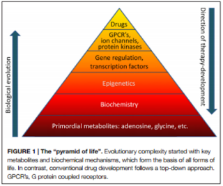
     Epilepsy is one of the most prevalent neurological condition characterized by spontaneously non-provoked seizures that occur as a result of a complex disorder of network homeostasis. Current treatments focus on the suppression of these epileptic seizures. However, research has come across evidence for the prevention of epileptogenesis (development and progression of epilepsy) through biochemical manipulations. Detlev Boison, in his review, The Biochemistry and Epigenetics of Epilepsy: Focus on Adenosine and Glycine has discussed this concept via the mechanisms implicated in epileptogenesis and the biochemical interactions between adenosine and glycine which serve as mayor contributors to the development of epilepsy.


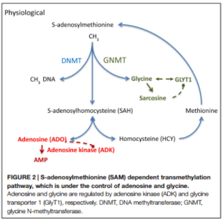
     Boisin focuses on temporal lobe epilepsy (TLE), especially its key metabolites adenosine and glycine.  These are primitive biological elements with important biochemical functions, whose homeostasis is generally affected in epileptic brains. Adenosine is an endogenous anticonvulsant and seizure terminator only when the adenosine A1 receptors is activated. Overexpression of its kinase (ADK), results in an adenosine deficiency associated with the increase of astrocytes, known as astrogliosis, and the adenosine receptor (AR) has been linked to the control of DNA methylation under the activity of ADK. The latter is expressed in both cytoplasmic and nuclear isoforms whose functions range from homeostatic regulation to modification in DNA methylation statuses. Astrogliosis is closely related with the increase in ADK expression and the deficiency of adenosine, which leads to the production of seizures and the hypermethylation of DNA. Therefore, it can be concluded that the dysregulation of ADK has a significant effect in the process of turning a normal brain into an epileptic one. Glycine, on the other hand, may have various effects depending on the activation of its presynaptic or postsynaptic receptor (GlyR’s). Low concentrations of glycine have pro-convulsive effects, while high concentrations reduce its occurrence. Its homeostasis is crucial in maintaining a balance in neuronal excitability and its regulation and reuptake is achieved by the glycine transporter 1 (GlyT1). The latter, when increased, is associated with TLE and has been considered a promising target for treatments of cognitive diseases.  

     “The knowledge of epigenetic mechanisms implicated in the development of epilepsy provides a conceptual and mechanistic framework for the future development of epigenetic therapies tailored to prevent epilepsy (antiepileptogenic) or its progression (disease modifying)”(Boison, 2016). The current treatments fail to take into account the causes of epilepsy and are therefore unable to halt epileptogenesis, which is why epigenetic modifications offer new therapeutic alternatives. Using rat models it has been discovered that an adenosine augmentation can effectively reduce and even suppress the occurrence of seizures. They have also been used to form the basis for both the ADK hypothesis (acute insults to the brain such as traumatic brain injury, seizures, or a stroke lead to an acute surge in adenosine associated with transient downregulation of ADK) and the methylation hypothesis of epileptogenesis (suggests that seizures may induce epigenetic modifications aggravating the condition). Alteration in DNA methylation plays a significant role in in the development and progression of neurodegenerative diseases like epilepsy. The increased activity of DNA methylating enzymes and the hypermethylation of DNA has been linked to onset of epilepsy. However, the status of DNA methylation depends on the equilibrium of biochemical enzyme reactions catalyzed by DNA methyltransferases (DNMTs) or Ten-eleven translocations (TET) enzymes. These mechanisms depend on transmethylation pathways controlled by adenosine and glycine concentrations regulated by ADK and GlyT1.


     The biochemical discoveries discussed by Boison have made way for new research areas. Understanding the epigenetics behind epilepsy may be result in the development of novel and effective therapeutic strategies.
 “Challenges for drug development remain. It needs to be determined whether new therapeutic agents can enter the brain and whether a higher level of selectivity for specific isoforms of ADK can be achieved. Due to the different distribution of nucleoside transporters within the brain there might be opportunities for the development of cell-type or isoform selective therapies.” (Boison, 2016).


References:
Boison, D. (2016). The Biochemistry and Epigenetics of Epilepsy: Focus on Adenosine and Glycine. Retrieved from https://www.ncbi.nlm.nih.gov/pmc/articles/PMC4829603/pdf/fnmol-09-00026.pdf

Saturday, November 12, 2016

Diving deeper: some epigenetic changes in epilepsy

The establishment and maintenance of epigenetic marks are crucial for normal development and function. Here, we will discuss how DNA methylation patterns are altered, and the presence od certain histone variants and microRNA's is changed in epilepsy. 

DNA Methylation

There is evidence in recent studies in animal models and tissues from epileptic patients that have revealed altered methylation patterns in DNA in these patients, compared to healthy individuals. 

An autopsy study of the hippocampus tissue from patients with temporal lobe epilepsy found a greater degree of methylation of the reelin promoter among the patients with epilepsy compared to controls (Kobow et al., 2009). Reelin is an extracellular matrix protein that performs key functions in neuronal migration, synaptic plasticity, and maintenance of the laminar structure of hippocampal granule cells. Loss of this structure in the hippocampal dentate nucleus (granule dispersion) is present in up to 50% of the patients affected by temporal lobe sclerosis. 

Additionally, another study examined DNMT1 and DNMT3A (DNA methyltransferase 1 and 3A) expression in patients with temporal lobe epilepsy compared to healthy controls. Remember that DNMT1 is responsible for the maintenance of methylation patterns, and DNMT3a and DNMT3b were involved in de novo methylations. The study concluded that both DNMT’s are more abundant in patients with temporal lobe epilepsy, hinting that they may contribute to the pathogenesis of this type of epilepsy (Zhu et al., 2011).

Finally, a study analyzed the overall DNA methylation in the hippocampus of rats with chronic epilepsy compared with controls. The group with chronic epilepsy showed more overall methylation (Kobow et al., 2013). But when provided with a ketogenic diet, they observed a reduction in seizure frequency and a change in the DNA methylation pattern.


Histone modification

Studies in animal models have demonstrated changes in chromatin mediated by histone modifications following epileptic seizures. 

For example, a study analyzed rat hippocampal tissue 3 hours after having induced status epilepticus and found that histone H4 hypoacetylation (which is a marker for gene repression) in the promoter of the glutamate 2 receptor (GluR2), as well as hyperacetylation (a marker for gene transcription) in the brain-derived neurotrophic factor (BDNF) promoter (Huang et al., 2002). These findings clearly show that status epilepticus* rapidly triggers modulations in histone acetylation. The same study found that prior administration of an HDAC (histone deacetylase) inhibitor prevented hyperacetylation of the GluR2 promoter, which could help design a treatment for epilepsy. 

In a more recent study, the same author reported greater HDAC2 expression in tissue from patients with temporal lobe epilepsy, as well as from animal subjects with status epilepticus, than in controls (Huang et al., 2011). HDAC2 is a type of HDAC expressed by the central nervous system that is active in neurodevelopment. Results from the study show that HDAC2 is significantly involved in the pathogenesis of temporal lobe epilepsy, and in the cognitive impairment that may sometimes also be associated with this type of epilepsy.

In another animal model of epilepsy using electrically induced seizures, Tsankova et al. found changes in the acetylation of histones H3 and H4 at the CREB promote region in the rat hippocampus, with H4 hypoacetylation of CREB and H3 hyperacetylation of CREB miRNA (Tsankova et al., 2004). CREB is an important transcriptional factor that plays an important role in the epileptogenic process. 


Micro-RNA and epilepsy

Several studies of the expression profile of miRNA in epilepsy have been published recently, and they offer promising information about the potential role of miRNA as a biomarker.

One example is a study that describes the miRNA expression profile in rats with induced status epilepticus based on analyses of brain tissue and blood samples (Liu et al., 2009). The authors found similar expression profiles for one miRNA subtype in blood and hippocampal tissue and therefore support the possibility that miRNA's might serve as blood biomarkers for epilepsy.

On the other hand, studies of miRNA are also providing additional knowledge about the epileptogenic process.

For example, various studies carried out in animal models have all shown increased expression of miRNA-132 in the hippocampus of rats with induced status epilepticus  (Pulido et al., 2015). It is understood that miRNA-132 has anti-inflammatory functions, and inflammation has been shown to play a role in epileptogenesis. Therefore, increased miRNA-132 may contribute to the development of epilepsy.


*
Status epilepticus is said to occur when a seizure lasts too long or when seizures occur close together and the person doesn't recover between seizures. 

More information can be found in this paper:


Pulido Fontes, L., Quesada Jimenez, P., & Mendioroz Iriarte, M. (2015). Epigenetics and epilepsy. Neurología (English Edition), 30(2), 111-118. http://dx.doi.org/10.1016/j.nrleng.2014.03.002